 ESPCI web site
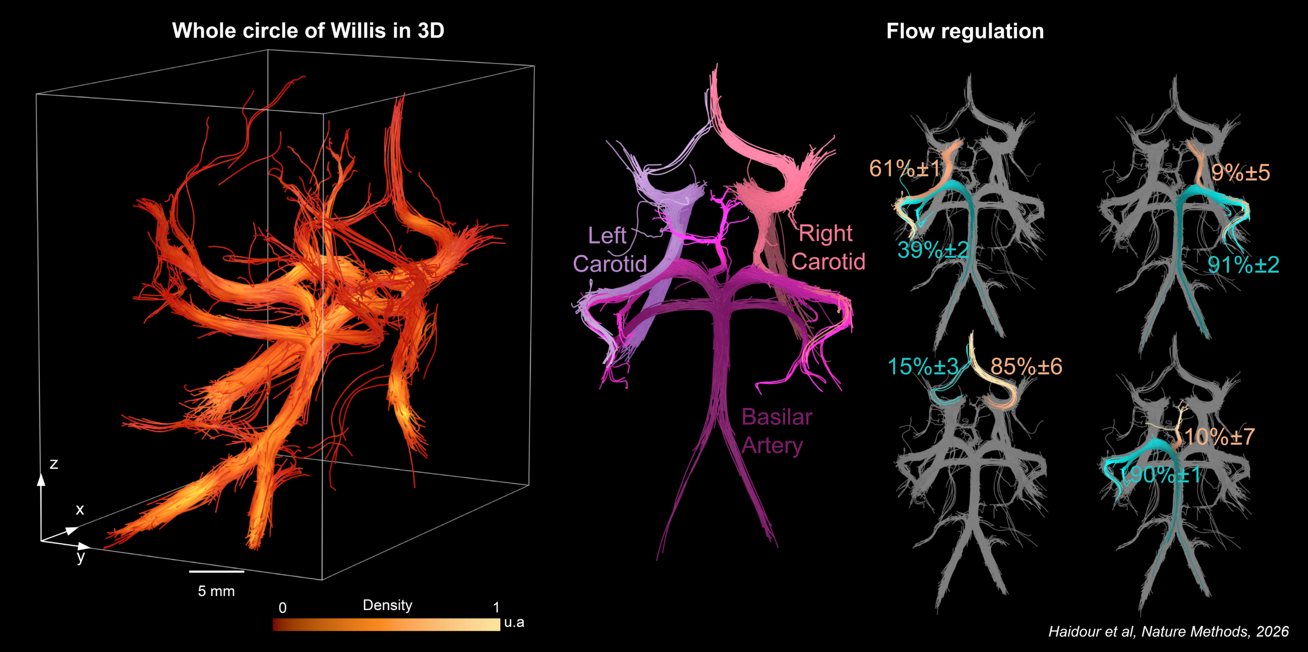
Imaging the Circle of Willis like never before: 4D Ultrasound Cerebral Angiography is now published in Nature Methods
We are proud to share that our former PhD student Nabil Haidour and our researcher Clément Papadacci introduced 4D Ultrasound Cerebral Angiography (4D UCA), a non-invasive technique that images the Circle of Willis, the ring of arteries at the base of the brain that reroutes blood when a carotid or vertebral artery is blocked, and quantifies its hemodynamics in 3D.
Until now, observing both the smallest arteries of this network and the blood flowing through them, across the skull, was out of reach. In primates, the team was able to image the entire Circle of Willis at depths of up to 7 cm, including fine vessels that reference MR angiography could not always detect. They also measured blood velocity, direction and flow partitioning at bifurcations, with a resolution of about 150 µm, and revealed marked differences in blood flow organization from one animal to the other. All of this without ionizing radiation, using an ultrasound contrast agent already approved for clinical use.
Behind this result: a custom multi-lens matrix probe, real-time optimization of ultrasound transmission through the skull, 3D ultrafast imaging, and tracking of individual microbubbles flowing in the blood.
This preclinical study opens the way to a better understanding of how the brain protects itself against stroke, and ultimately to new brain imaging tools for humans.
Congratulations to Nabil, Clément, and all co-authors at Institute Physics for Medicine and the Paris Brain Institute (ICM), and thanks to the ERC Starting Grant MicroFlowLife for supporting this work.
📄 Read the paper: www.nature.com/articles/s41592-026-03314-4
🎥 3D view of the Circle of Willis reconstructed from the trajectories of individual microbubbles by Alexandre Dizeux


