 ESPCI web site
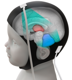
Design of optimal wearable ultrasound probe for neonatal functional ultrasound imaging using an in silico realistic brain phantom and in vitro validation.
Project description :
Objective of the project: determine the characteristics of an optimal ultrasound imaging (sensor and imaging mode) for functional ultrasound brain monitoring in the neonate.
Context: Recent work at Physique pour la Médecine Paris has opened major new possibilities for using ultrasound in neuroscience. We developed a new brain imaging method called functional Ultrasound (fUltrasound), similar in principle to fMRI, which can visualize brain activity while it responds to stimuli or performs tasks. The technique relies on ultrafast imaging of blood flow, making it highly sensitive to small changes in cerebral blood volume and able to detect neurovascular coupling (increased blood flow linked to local neuron activation).
One promising clinical application is functional brain imaging in newborns through the fontanelle. This approach has already allowed the detection of epileptic foci in newborns with brain disorders—children who often resist treatment and for whom conventional imaging options are limited. Beyond that, the method could be used to study brain connectivity for diagnostics, or to investigate fundamental questions about development.
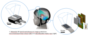
Goal and methods: To optimize the ultrasound probes used for neonatal fUS imaging, researchers rely on computer simulations of ultrasound propagation through the newborn brain via the fontanelle. These simulations help determine the best probe size and geometry for this unique entry point, which can vary significantly between infants. The student’s task will be to create a realistic model of the brain, skull, and fontanelle as an acoustic medium, then to simulate ultrasound propagation with different probe designs (linear array, µbeamformed matrix array) and quantify relevant readouts (sensitivity, volume coverage). From these tests, they will identify the most suitable configurations for developing future neonatal fUS probes. The student will also be able to validate the imaging modes on realistic neonate phantoms, with real micromotorized and µbeamformed probes, using an ultrafast imaging ultrasound platform.
Candidate Profile: We are looking for a student who is interested in the interface between physics and medicine, proficient with coding (python or matlab), preferably with some experience in benchtop experiment.
Supervisor:
Name: Charlie Demené and Jérome Baranger
Contact e-mail: charlie.demene@espci.fr, jerome.baranger@espci.fr


