 ESPCI web site
fUS imaging becomes compatible with hybrid PET/CT/fUS Imaging
Currently boiling, imaging medical might have found an unprecedented technique for observing multiple facets of living in real time and non-invasively. Indeed, our team at ESPCI Paris – University PSL (Inserm/CNRS/ESPCI Paris), our Research Accelerator Technological (A.R.T. Inserm) in Biomedical Ultrasound and the research center cardiovascular clinic (Inserm / University Paris Descartes) have developed a new medical imaging instrument associating positron emission tomography -“Pet-scan” – with ultrafast ultrasound imaging, christened PETRUS for Positron Emission Tomography Registered Ultrafast Sonography. They obtained images in three dimensions where anatomy, metabolism, the functionality and even the elasticity of organs are perfectly superimposed. These works are the cover of the journal Nature Biomedical Engineering on February 6th.
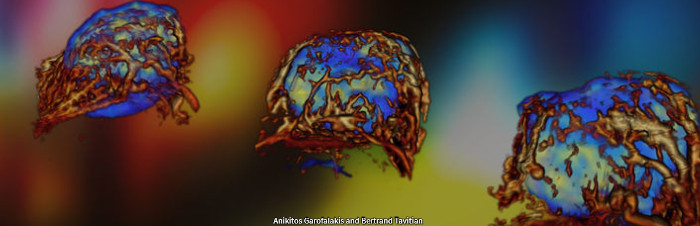
Funded on this project by France Life Imaging organization, our colleagues from PARCC Inserm and our team were able to test our method from marketed instruments and assembled without major modification. they have especially imaged cancerous tumors in mice, or cardiac activity in the rat to test the timing of the two methods and the complementarity of parameters observed. Because it allows view multiple parameters simultaneously basic biology in the form of maps parametric quantitative, this new imaging reflects even more finely the complexity of the topology of the living. This totally traumatic technique real time living observation offersmany perspectives: exploring the link between organ metabolism and vascularization like the heart, kidney or liver, follow more precisely the effect of the new cancer treatments, characterize the follow-up infarction, etc. Developed in a context preclinical, PETRUS is a technology high performance easily translatable clinically for biomedical research.
For neuroscience, these results demonstrate that fUS imaging developed in ERC project FUSIMAGINE is fully compatible with PET/CT hybrid imaging, in order to give access simultaneously to anatomy, metabolism and neurofunctional imaging.
For more information :
Provost J et al. Simultaneous positron emission tomography and ultrafast ultrasound for hybrid molecular, anatomical and functional imaging, Nature Biomedl Eng 2018; 2(2): 85-94, doi:10.1038/s41551-018-0188-z
Contact researchers :
Bertrand Tavitian, bertrand.tavitian@inserm.fr
Mickaël Tanter, mickael.tanter@espci.fr


