Imaging the brain through the skull: a comprehensive review
A new perspective in Nature Biomedical Engineering reviews the complexity of imaging the brain with ultrasound and optical waves. The article highlights current advantages and limitations of each modality, and discusses innovative strategies to correct skull-induced distortions in ultrasound, optical, and hybrid acousto-optic imaging.
Understanding the human brain is one of the greatest challenges in science and medicine. While MRI and CT scans are widely used, techniques based on light and ultrasound offer unique advantages: they can deliver high resolution, lower costs, and even be used at the bedside. Yet one obstacle stands in the way—the skull.
The skull is a powerful barrier: it scatters and absorbs light, while also weakening and distorting ultrasound waves. This makes non-invasive brain imaging extremely difficult, especially in adults.
Light-based imaging
Optical methods, particularly those using near-infrared light, can visualize brain activity in animals and newborns. However, in adults most light is blocked by the skull. Researchers are now designing new light-sensitive agents and advanced computer models to improve resolution and penetration.
Ultrasound imaging
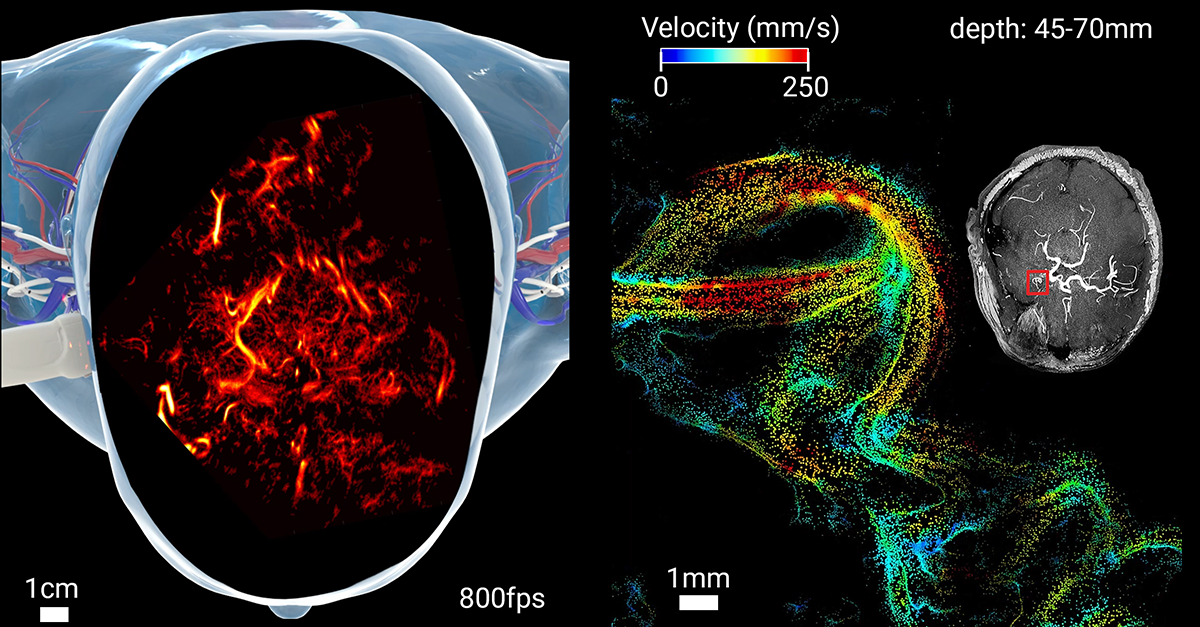
Ultrasound is already used to measure blood flow in the brain, especially in infants where the skull is thinner. Recent breakthroughs such as ultrasound localization microscopy, which tracks tiny contrast microbubbles, have achieved unprecedented detail in both animal studies and first human applications.
Hybrid approaches: light meets sound
Optoacoustic imaging combines light and ultrasound, capturing high-resolution images of brain activity without the invasive procedures traditionally required. Early studies in rodents and patients show promising results, despite the technical challenges posed by the skull.
Looking ahead
By refining correction techniques and merging imaging modalities, researchers are gradually overcoming the skull’s distortions. These advances pave the way toward safe, non-invasive, and high-resolution imaging of the living brain, with potential to transform both neuroscience research and clinical care.
👉 Read the full paper: https://doi.org/10.1038/s41551-025-01433-5
📷 Transcranial ULM in humans boosts the spatial resolution performance of pulse-echo ultrasound by almost two orders of magnitude (left) while providing non-invasive measurements of cerebral haemodynamics and quantified readings in small vessels (<100 μm in diameter) embedded at depths of several centimetres in the human brain (right).


