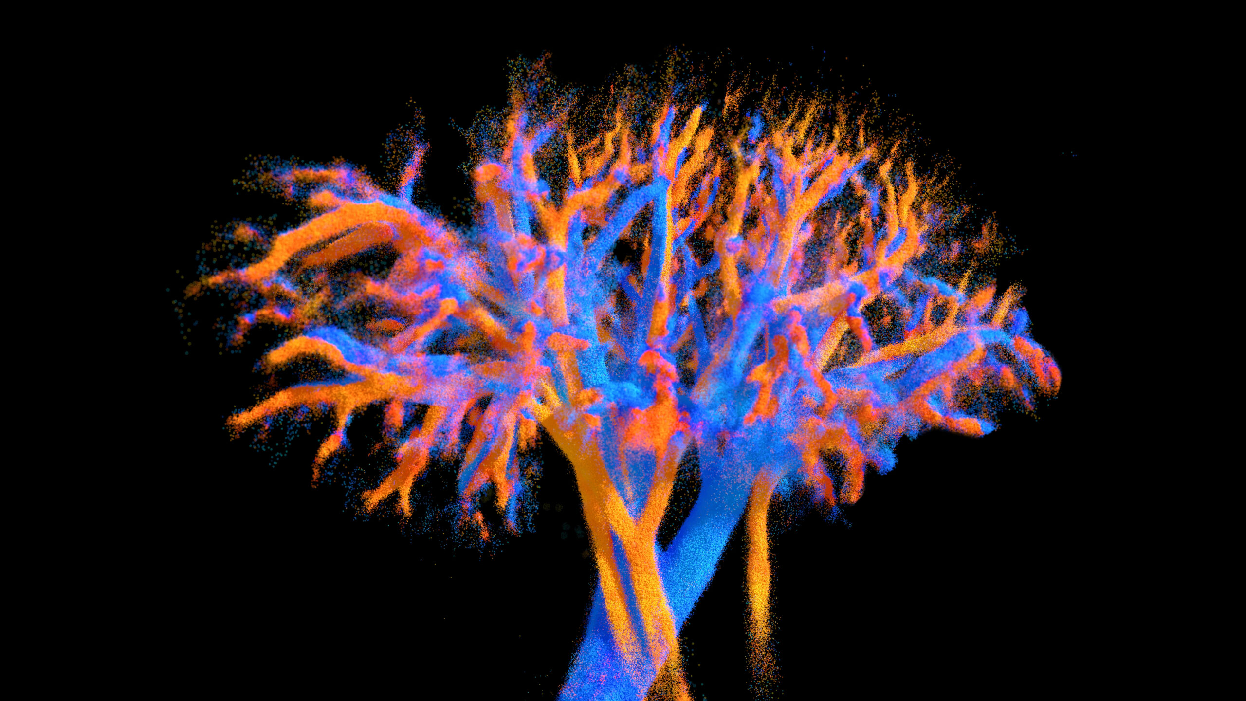
Major breakthrough in medical imaging just published in Nature Communications
Researchers develop an ultrasound probe capable of visualising an entire organ in 4D
For the first time, whole-organ blood flow dynamics can be mapped at micrometric resolution using an ultrasound multi-lens probe — demonstrated in vivo in the heart, kidney, and liver.
Developed at Physics for Medicine Paris, this technology enables real-time 4D imaging of vascular dynamics across entire organs.
A probe inspired by optical lens design
The innovation relies on a multi-lens ultrasound probe (8 × 10 cm, 252 elements), each element equipped with a tiny acoustic lens. This design, inspired by optics, enables an unprecedented combination of wide coverage and high resolution — two features that had been mutually exclusive until now.
The probe achieves:
- imaging volumes up to 120 × 100 × 82 mm³,
- a spatial resolution around 100 µm,
- and ultrafast acquisition at 312 volumes per second.
These performances allow the visualisation of both large vessels and the finest microcirculation, revealing the complex vascular networks that sustain tissue function.
A step toward non-invasive vascular imaging
The study, carried out as part of the ERC Starting Grant MicroFlowLife, was led by Nabil Haidour under the supervision of Clément Papadacci, Inserm researcher at Physics for Medicine Paris.
It was conducted in collaboration with a team from IMRB – Institut Mondor de Recherche Biomédicale and Hôpital Henri Mondor (AP-HP, Créteil).
By mapping blood flow in full 4D across large organs, this technology represents a key advance in ultrasound imaging — bridging the gap between large-scale anatomical imaging and fine vascular resolution.
It opens new possibilities for studying microcirculation and diagnosing small vessel diseases, which remain challenging to detect with current imaging methods.
From preclinical imaging to the clinic
The technology will now be tested in humans as part of an upcoming clinical trial.
Further developments for clinical deployment are being carried out with the support of the Technological Research Accelerator for Biomedical Ultrasound (ART Ultrasons Biomédicaux), created by Inserm and integrated into Physics for Medicine Paris.
“The probe can be connected to small portable equipment, which would allow its integration into clinical practice,” explains Clément Papadacci.
By offering comprehensive and dynamic visualisation of the vascular system, this approach could become a powerful tool for understanding blood flow regulation, diagnosing microcirculation disorders, and monitoring treatment response.
Reference and media
📄 Read the full paper : Multi-lens ultrasound arrays enable large scale three-dimensional micro-vascularization characterization over whole organs – Nature Communications, October 28, 2025
🔗 https://doi.org/10.1038/s41467-025-64911-z
Inserm press release in French
Inserm press release in English


