 ESPCI web site

Research Internship – Non-invasive pressure sensing using Ultrasound Localization Microscopy
Scientific Context: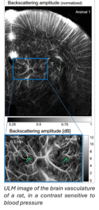
Pressure is a critical physiological parameter that plays a key role in diagnosing and monitoring various medical conditions. For example, measuring pressure within the left ventricle helps detect cardiac abnormalities, while monitoring pressure in the portal vein can aid in diagnosing hypertension. Similarly, intracranial pressure measurements are essential after brain trauma to prevent further tissue damage. Currently, the gold standard for pressure measurement remains invasive techniques, such as introducing a catheter into the vasculature or heart, or using small pressure probes in the brain through a burr hole in the skull.
Ultrasound Localization Microscopy (ULM) is a cutting-edge technique that uses micro-bubbles as contrast agents. These micro-bubbles, injected into the vasculature, are localized and tracked through ultrasound imaging, enabling the creation of high-resolution maps of hemodynamic properties (see Figure) [1].
Recent studies have shown that these micro-bubbles are also sensitive to the hydrostatic pressure in their environment, suggesting their potential as non-invasive sensors for pressure monitoring throughout the body. Our preliminary results, however, are impaired by the polydispersity of the microbubbles available for clinical use, as well as by the variability inherent to in vivo imaging.
Objectives of the project:
Develop an in vitro setup and data analysis pipeline for pressure monitoring using ultrasound imaging and microbubbles.
Methods and internship organisation:
In this internship, the student will develop an in vitro setup to image flowing microbubbles in well-controlled pressure conditions. Once this setup is operational, the objective is to test different imaging systems (for e.g,. varying ultrasound frequencies) as well as different contrast agents (for e.g., polydisperse or monodisperse microbubbles). Based on these results, we will try to build a physical model of the microbubble response and identify optimal conditions for pressure monitoring.
Specifically, the student will:
- Build an in vitro setup for ULM data acquisition in varying pressure conditions.
- Acquire and analyze ULM data on this setup
- Build a physical model for the microbubble response.
Candidate profile:
We are looking for a student who is interested in the interface between physics and medicine, preferably with some experience in benchtop experiments.
Duration: 6 months
Location: Institute Physics for Medicine Paris, 2-10 rue d’Oradour-sur-Glane, 75015 Paris
Contact: justine.robin@espci.fr, charlie.demene@espci.fr
[1] C. Errico et al., « Ultrafast ultrasound localization microscopy for deep super-resolution vascular imaging », Nature, vol. 527, no 7579, p. 499‑502, nov. 2015, doi: 10.1038/nature16066.


