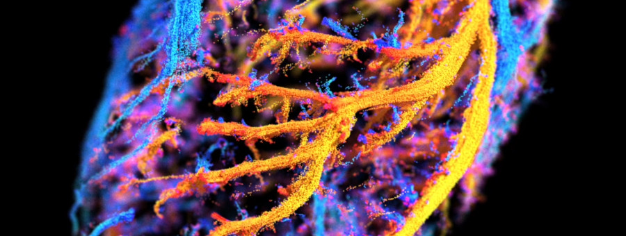
The heart microcirculation captured in 3D using ultrasound microscopy
Our recent publication in JACC Cardiovascular Imaging shows the coronary microvasculature captured in the beating heart, in 3D with a spatial resolution of 20 microns. The data were collected using 3D ultrafast ultrasound localization microscopy, and carry unvaluable information to assess both the coronary anatomy and function.
The heart muscle, called the myocardium, is supplied in blood by a network of vessels with diameters varying from a few millimeters down to a few microns. The anatomy of largest coronary arteries can be seen with current imaging modalities, but there is no tool to directly image and measure the blood circulation in the microvasculature. Yet, this coronary microcirculation, comprising of vessels smaller than 300 microns, plays a major role in the control of the heart perfusion: coronary microvascular diseases are associated with a high rate of heart failure, cardiac arrest, or myocardial infarction.
Ultrasound localization microscopy (ULM) is capable of imaging non-invasively the blood flows across several spatial scales: micron-scale vessels can be imaged at several centimeters deep in organs. Our laboratory Physics for Medicine, and more specifically the research team of Mathieu Pernot and his PhD student Oscar Demeulenaere, has developed 3D ultrafast ULM to capture the coronary microcirculation across the entire heart.
The experiments demonstrate the feasibility of 3D coronary ULM in vivo in rodents. Ultrafast ultrasound can be translated to clinics as a non-invasive, non-ionizing, and portable imaging technology. 3D coronary ULM is therefore expected to become a powerful tool for clinical investigation of microvascular diseases, not only in the field of cardiology but also for addressing brain or liver diseases.
Video credits: Alexandre Dizeux / Physics for Medicine Paris
Full publication: Demeulenaere O, Sandoval Z, Mateo P, Dizeux A, Villemain O, Gallet R, Ghaleh B, Deffieux T, Demené C, Tanter M, Papadacci C, Pernot M, Coronary flow assessment using 3-dimensional ultrafast ultrasound localization microscopy, JACC: Cardiovascular Imaging, 2022, https://doi.org/10.1016/j.jcmg.2022.02.008


