 ESPCI web site
The New Wave of Cardiovascular Biomechanics
Our recent work on cardiovascular imaging has been highlighted in the editorial of JACC: Cardiovascular Imaging as part of “the new wave of cardiovascular biomechanics”. In this editorial, Prof. Sengupta and Chandrashekhar point out the need for non-invasive methods to quantify cardiac biomaterial properties in vivo, and particularly to assess the cardiac structure-function relationship in clinical settings. Six articles published by Mathieu Pernot and his team are cited as significant advances “creating unprecedented opportunities in positioning cardiac imagers at the heart of precision medicine“. The editorial further explains the transformation of patient care that can be expected from the combination of advanced cardiac imaging methods, computational modelling, machine learning and biomaterial design.
Fortunately, the technological advances in cardiac imaging have provided newer opportunities for applying the engineering toolbox in clinical practice, and iJACC has had the opportunity to showcase some of these important developments. One of the most exciting is ultrafast echocardiography, which images with high temporal resolution (many thousands of frames per second) and offers unique opportunities in measuring cardiac material properties (e.g., stiffness), as well as functional data such as electromechanical activation mapping and temporally enhanced blood flow imaging.
P.P. Sengupta & Y. Chandrashekhar
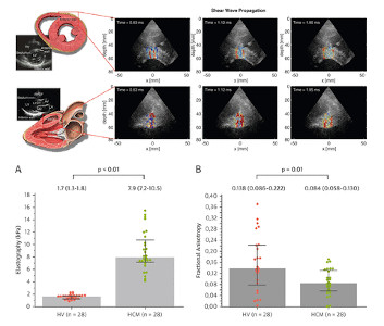
Over the past ten years, Mathieu Pernot and his team have successfully implemented ultrafast ultrasound imaging for clinical cardiovascular applications. In particular, they have demonstrated the robustness of shear wave elastography to measure the myocardial stiffness in adults and infants and differentiate patients with hypertophic cardiomyopathy from healthy volunteers (Villemain et al., JACC Cardiovascular Imaging 2018; Villemain et al., JACC Cardiovascular Imaging 2019). They have also provided ultrasound-based methods to non-invasively map the myocardial fiber architecture and its dynamics during a cardiac cycle, providing new means to assess the link between structure and function (Papadacci et al., Science Report 2017; Lee et al., Physics for Medicine and Biology 2012). In a recent study, the team has also leveraged the exquisite sensivity of ultrafast Doppler imaging to image the coronary vasculature in healthy volunteers (Maresca et al., JACC Cardiovascular Imaging 2018). Ultrasound-based coronary imaging is expected to have a huge impact for the diagnosis of microcirculation and microvascular disease, as it will provide a performant and non-invasive alternative to X-ray coronarography.
- Villemain O, Correia M, Mousseaux E, Baranger J, Zarka S, Podetti I, et al. Myocardial Stiffness Evaluation Using Noninvasive Shear Wave Imaging in Healthy and Hypertrophic Cardiomyopathic Adults. JACC: Cardiovascular Imaging 2018. https://doi.org/10.1016/j.jcmg.2018.02.002.
- Villemain O, Correia M, Khraiche D, Podetti I, Meot M, Legendre A, et al. Myocardial Stiffness Assessment Using Shear Wave Imaging in Pediatric Hypertrophic Cardiomyopathy. JACC: Cardiovascular Imaging 2018;11:779–81. https://doi.org/10.1016/j.jcmg.2017.08.018.
- Maresca D, Correia M, Villemain O, Bizé A, Sambin L, Tanter M, et al. Noninvasive Imaging of the Coronary Vasculature Using Ultrafast Ultrasound. JACC: Cardiovascular Imaging 2018;11:798–808. https://doi.org/10.1016/j.jcmg.2017.05.021.
- Villemain O, Sitefane F, Pernot M, Malekzadeh-Milani S, Tanter M, Bonnet D, et al. Toward Noninvasive Assessment of CVP Variations Using Real-Time and Quantitative Liver Stiffness Estimation. JACC: Cardiovascular Imaging 2017;10:1285–6. https://doi.org/10.1016/j.jcmg.2017.01.018.
- Papadacci C, Finel V, Provost J, Villemain O, Bruneval P, Gennisson J-L, et al. Imaging the dynamics of cardiac fiber orientation in vivo using 3D Ultrasound Backscatter Tensor Imaging. Scientific Reports 2017;7:. https://doi.org/10.1038/s41598-017-00946-7.
- Lee W-N, Larrat B, Pernot M, Tanter M. Ultrasound elastic tensor imaging: Comparison with MR diffusion tensor imaging in the myocardium. Physics in Medicine and Biology 2012;57:5075–95. https://doi.org/10.1088/0031-9155/57/16/5075.


