 ESPCI web site

Ultrasound angiography of coronaries goes 3D
A recent publication of our group, published in Physics in Medicine and Biology, reports the first implementation in 3D of ultrafast Doppler for coronary flows imaging in animal models. The results were advertised in Physics World in an article emphasizing the technical achievements as well as the clinical potential of the method.
Coronary arteries are responsible for supplying oxygenated blood to the heart muscle (called myocardium) and perturbations of coronary blood flows have deleterious effects on the heart function. The associated pathologies include angina pectoris, myocardial infection, or myocardial infarct. Clinically available techniques to assess the coronary blood flows are either invasive (invasive coronary angiography), ionizing (FFR-CT, fractional flow reserve using computed-tomography), and/or limited to the visualization of large coronary arteries (MRI, magnetic resonance imaging). Our laboratory Physics for Medicine Paris proposes ultrafast ultrasound as a non-invasive and highly sensitive alternative for imaging coronary arteries. This new method was coined Ultrafast Doppler Coronary Angiography (UDCA) and provides quantification of the coronary blood flows.
In 2018, the team of Mathieu Pernot demonstrated UDCA in two-dimension in the beating heart of animal models and patients. Expanding UDCA to three-dimension imaging not only enables to capture the complex architecture of coronary arteries, but also to quantify the absolute coronary flow rate. Such quantification was not achievable in 2D, as the flow rate estimate depends on the orientation of the artery in the imaging plane.
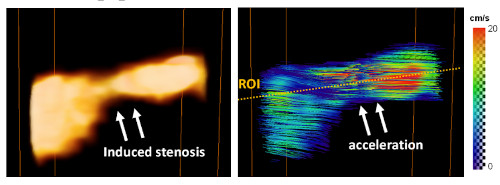
For the first time, coronary flows were imaged and quantified in 3D in swine having a partial occlusion (stenosis) of the left anterior descending artery. As expected, a reduced Power-Doppler signal (signature of a reduced diameter) as well as an acceleration of the flow were observed in the stenosis region (see figure below). Measurements were validated through comparison with an invasive flowmeter placed at the vicinity of the imaged region.
This preliminary validation study was performed in open-chest animals to ensure optimal imaging conditions. Implementation of a fully non-invasive (i.e trans-thoracic or trans-oesophagial) UDCA is a key requirement for clinical translation. Besides, the field-of-view must also be extended to cover the entire heart. Mathieu Pernot and his team are working on addressing these issues to develop UDCA for human use. The method could have a major impact in the diagnosis of microvascular coronary diseases as well as in the planning of coronary surgeries.
Full citation:
Correia M, Maresca D, Goudot G, Villemain O, Bizé A, Sambin L, et al. Quantitative imaging of coronary flows using 3D ultrafast Doppler coronary angiography. Phys Med Biol 2020;65:105013. https://doi.org/10.1088/1361-6560/ab8d78.
References:
Maresca D, Correia M, Villemain O, Bize A, Sambin L, Tanter M, et al. Noninvasive Imaging of the Coronary Vasculature Using Ultrafast Ultrasound. JACC-Cardiovascular Imaging 2018;11:798–808. https://doi.org/10.1016/j.jcmg.2017.05.021. Maresca D, Correia M, Tanter M, Ghaleh B, Pernot M. Adaptive Spatiotemporal Filtering for Coronary Ultrafast Doppler Angiography. IEEE Transactions on Ultrasonics Ferroelectrics and Frequency Control 2018;65:2201–4. https://doi.org/10.1109/TUFFC.2018.2870083


